

Emma Wilson, associate professor of biomedical sciences, along with several other UCR School of Medicine researchers, have recently uncovered the effects of a parasitic disease, toxoplasmosis, on cells in the brain as well as the mechanism by which these effects manifest. By understanding the influence of this parasite on brain tissue, Wilson and fellow researchers hope to better the treatment of this disease and ultimately develop therapies that can aid individuals afflicted with different neurological diseases.
The parasite that causes toxoplasmosis, toxoplasma gondii, is found around the world, and according to the Center for Disease Control (CDC), it affects about 60 million Americans. The most common way people acquire the parasite is through the consumption of undercooked meat or by eating in unsanitary conditions. However, a majority of individuals that act as hosts for the parasite do not manifest symptoms because the parasite cannot exert its malignant effects on individuals with healthy immune systems. Individuals with compromised immune systems, as well as pregnant women, show an increased risk in displaying the deleterious effects of toxoplasmosis, such as initial flu-like symptoms, which can be followed by damage to the brain, eyes and other organs. In pregnant women, birth defects have also been seen to accompany infection of the parasite.
Wilson, in collaboration with other researchers, explained their motivation for studying this parasite in her recently published paper in the PLOS Pathogens journal. Wilson explained how previous research and literature has suggested that toxoplasma contributes to the development of Parkinson’s disease, Alzheimer’s disease and even schizophrenia, leading to the idea of a “potential relationship of infection with toxoplasmosa on neurodegenerative and psychiatric disorders.”
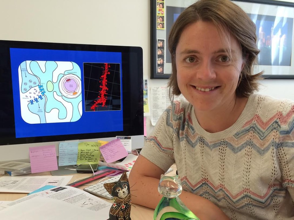
The results of the research showed that mice afflicted with toxoplasmosis displayed elevated levels of glutamate in the areas between neurons. Glutamate, a neurotransmitter or chemical, is normally released by a neuron that in turn causes the firing of surrounding neurons, and permits for vital cell to cell communication. It is imperative that this neurotransmitter be regulated and controlled in the brain because “unregulated levels can cause neuroexcitotoxicity,” which can lead to overactivation of the surrounding cells and subsequently, impaired brain function. The findings suggest that this buildup of glutamate can be attributed to dysfunctional astrocytes, or surrounding support cells of the brain, that cease to reuptake the glutamate.
The mechanism suggested by Wilson’s experiments, namely the buildup of glutamate in the area between neurons by the altered astrocytes, which seemingly exacerbates the effects of toxoplasmosis, was further corroborated by application of the antibiotic ceftriaxone. This antibiotic caused reactivation of the necessary proteins that are involved with glutamate control in astrocytes. In an interview with the Press-Enterprise, Wilson revealed that after the administration of ceftriaxone, the effects were qualitatively visible in affected mice, where their “erratic and risky behavior … returned to normal.”
Ultimately, Wilson is looking to delve deeper in the connection between toxoplasmosis and neurotransmitter level alterations with other neurodegenerative disorders. Additionally, Wilson wants to further investigate the specific cellular mechanisms by which toxoplasmosis initiates these astrocytes to stop the regulation of glutamate.


